AI adapts ultrasound to each patient
NVIDIA and Siemens Healthineers have presented `NV-Raw2Insights-US`, an AI system that analyzes raw ultrasound signals to estimate how sound travels through each patient and adjust focus in real time. The technology is still in the research phase and is not yet authorized for clinical or commercial use.
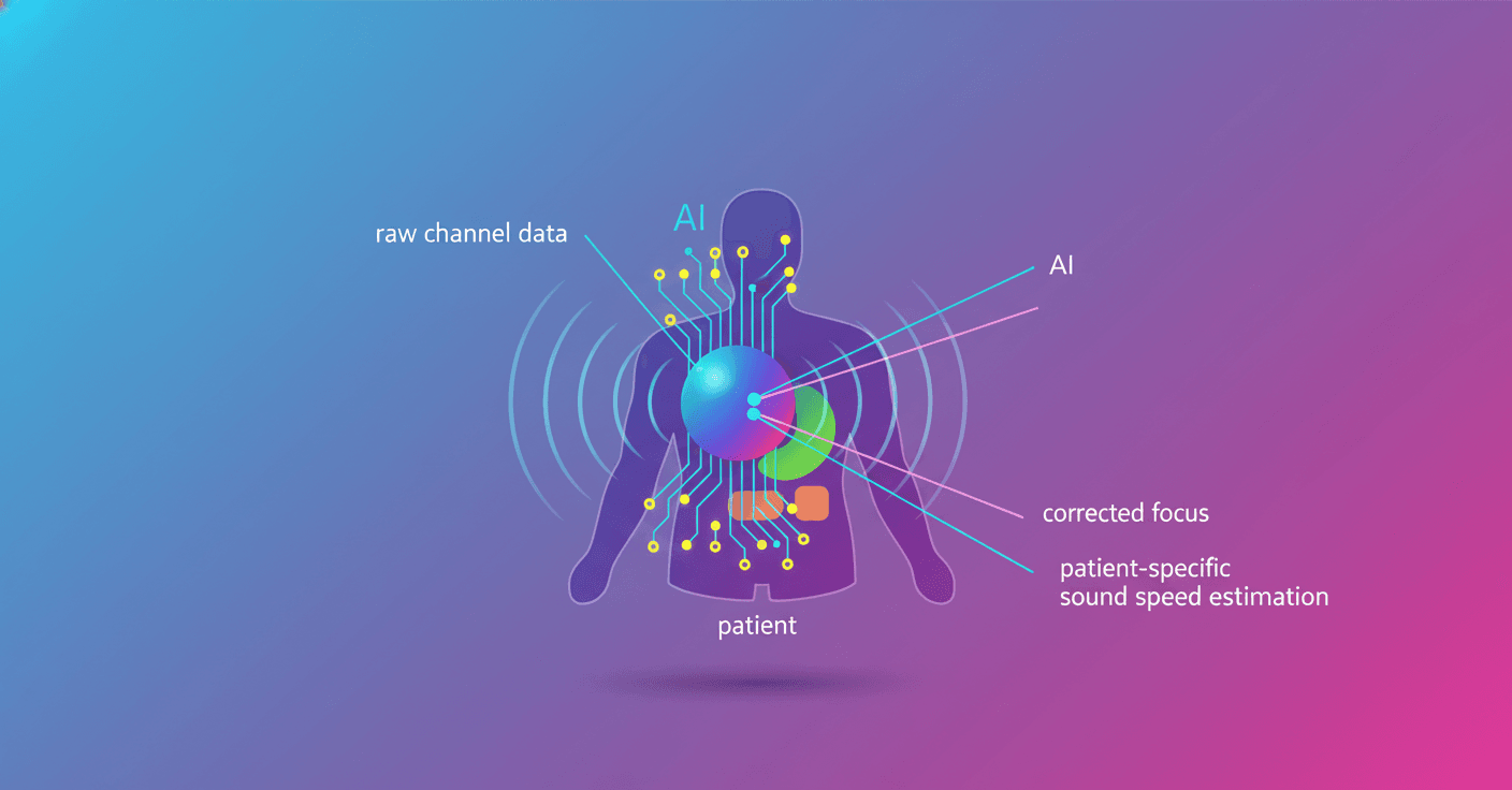
NVIDIA and Siemens Healthineers have presented NV-Raw2Insights-US, an AI model that analyzes the signals captured directly by an ultrasound probe to adjust image focus to each patient’s characteristics.
The system is still in the research phase and is not authorized for sale or clinical use in the United States or elsewhere. But it points to an important shift: treating ultrasound as more than a finished image and making use of the original data collected by the sensor.
Ultrasound does not start with an image
When you look at an ultrasound scan, the screen shows an image reconstructed from millions of tiny echoes bouncing through the body. Before reaching the monitor, that data goes through a process known as beamforming, which combines the signals and calculates where each echo appears to come from.
That process works, but it simplifies the physics. One common assumption is that sound travels at the same speed through all tissues. In reality, that speed changes depending on the part of the body it passes through. The result can be a less focused image or details that appear displaced.
NV-Raw2Insights-US attempts to correct that limitation. Instead of receiving an already processed image, it learns from the unreconstructed signals recorded by the probe. It uses them to estimate a personalized map of sound speed inside the patient and uses that map to improve focusing during the scan.
What changes in practice
In conventional ultrasound, adapting focus to the specific properties of each body requires complex calculations and may rely on broad assumptions. The model aims to make that adjustment in a single AI pass and send it back to the scanner while the image updates live.
That could be useful in situations where anatomy or tissue makes it difficult to obtain a clear image. For example, the system could account for the fact that sound behaves differently as it passes through different tissue layers, rather than applying one uniform speed to the entire body.
The difference is not only the use of a neural network. It is the starting point:
- Traditional systems work mainly with reconstructed images.
NV-Raw2Insights-USuses the original data from the probe’s channels.- The AI estimates the patient’s physical properties and uses them to adjust the image.
How AI connects to an existing ultrasound machine
Raw ultrasound data is large in volume and is not usually easy to access on clinical equipment. To address this, NVIDIA uses Holoscan Sensor Bridge, an open-source FPGA-based component that transfers the data to a GPU with low latency.
In the demonstration, an Altera Agilex-7 FPGA kit captures signals from an ACUSON Sequoia ultrasound system through its DisplayPort outputs. The data then travels over Ethernet to an NVIDIA IGX system, where the model runs inference on a GPU from the Blackwell family.
The sound-speed estimate then returns to the scanner to modify image focus live. NVIDIA deploys the system with Holoscan, a platform designed to process sensor data and run AI in real time on systems such as NVIDIA IGX Thor and DGX Spark.
What it does not mean yet
The demonstration is not the same as an available medical tool, nor does it prove on its own that the system improves diagnoses in hospitals. NVIDIA explicitly states that the technology is still under development and that future availability is not guaranteed.
What it does show is one possible path toward more adaptive ultrasound systems: models that not only interpret the final image, but also understand how it was generated and adjust the process for each patient. The next step will be determining whether that technical improvement leads to images that are more useful clinically and to more accurate medical decisions.